Reverse Phase - High Performance Liquid Chromatography (RP-HPLC)
RP-HPLC was used to:- determine the total amount of β-carotenes (2H-BC + non-2H-BC) and retinols (endogenous retinols + 13C10-retinols + 2H-retinols) circulating in the total serum of the subjects.
 |
| Set-up of Reverse-Phase HPLC |
2mL of chloroform, 1mL of methanol and 2mL of hexane were added to a 100uL serum sample. The mixture was centrifuged (3000rpm/10 minutes/ 4°C) so that the non-polar retinols and β-carotenes partition at the bottom together with the non-polar chloroform and hexane layers. The chloroform and hexane layers were combined and evaporated in inert nitrogen gas so that the retinols and β-carotenes materialised as white solid residues.
 |
| Nitrogen evaporator |
The solid residues of retinols and β-carotenes were re-dissolved completely in 100uL ethanol and 20uL of this mixture was injected into the RP-HPLC system.
Parameters
|
Description
|
Packed column
|
150mm length; 4.6mm internal diameter
particle size about 10um |
Stationary phase
|
C-30
organochlorosilanes coated around silica particles
(NON-POLAR)
|
Mobile phase
|
100% POLAR ethanol (initially)
|
Mode of operation
|
Column left under ambient conditions (without temperature control)
|
Gradient elution
Composition of mobile phase
changes progressively from high [ethanol] to low [ethanol] + high [another
non-polar solvent].
|
|
Detector
|
UV-Vis detector (fixed wavelengths)
|
Stationary phase
Non-polar β-carotenes, retinols and other carotenoids will interact with the non-polar C-30 stationary phase via van der Waal's forces and adsorb strongly against the stationary phase. If a polar stationary phase had been used instead, there would be little or no adsorption. The β-carotenes, retinols and carotenoids would be pushed along the column by the liquid mobile phase and be eluted quickly and very closely together, resulting in poor separation of β-carotenes and retinols.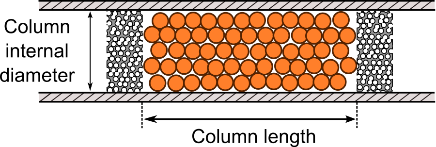 |
| Packed column in HPLC - stationary phase bonded to silica particles packed tightly in column |
 | |||
| Organochlorosilanes are chemically-bonded into silica particles |
Why was ethanol used as the liquid mobile phase/solvent?
Polar ethanol has a very weak eluotropic strength against a non-polar stationary phase. Thus at the beginning of the RP-HPLC operation, the non-polar retinols and carotenoids are not desorbed from the stationary phase; they are kept adsorbed onto the stationary phase for as long as possible so that other more polar analytes in the sample are eluted first, much earlier than the carotenoids and retinols. This allows for a clear separation of retinols and carotenoids from other impurities in the sample.Ethanol does not absorb at 340nm and 450nm and hence will not interfere with the detection of retinols and carotenoids in the serum samples. If ethanol had been able to absorb strongly at such wavelengths, the baseline would be very noisy and the signal-to-noise (S/N) ratio would be low. A real signal for the elution of carotenoids and retinols might be mistaken for part of the background noise and ignored. The HPLC system becomes much less sensitive.

Gradient elution
Initially, 100% polar ethanol with a very weak eluotropic strength was used as the mobile phase. As non-polar β-carotenes, retinols and other carotenoids adsorb strongly against the non-polar stationary phase, eluotropic strength of the mobile phase will have to be strengthened gradually (by adding more of non-polar solvents, such as hexane, and reducing the proportion of polar ethanol) so that the β-carotenes, retinols and other carotenoids can be eventually displaced from the stationary phase. This prevents long analysis times and band broadening.UV-Vis Photodiodearray detector : Fixed WVL
β-carotenes and carotenoids have extended conjugation and thus absorbs strongly at long wavelengths (450nm). Retinols do not have extended conjugation and thus absorbs strongly at shorter wavelengths (340nm).
The same sample is run twice separately with 20uL each - in the 1st run, the PDA detector is set at 340nm so that only retinol is detected and shows as a peak.
In the 2nd run, the PDA detector is set at 450nm so that only carotenoids (including β-carotenes) are detected. This produces multiple peaks in the chromatogram as there are many different carotenoids in the yellow maize. The peak for β-carotenes is identified by retention time matching.
 |
| Peaks for β-carotenes were identified by retention time matching |
Quantification of retinols and β-carotenes in total serum by EXTERNAL STANDARDS
A series of retinol standards of known concentrations were injected separately into the HPLC under the same chromatographic conditions as the serum sample. A calibration graph of area under the curve of retinol peak against concentration of retinol standard was plotted and a regression equation was derived.Area under curve of retinol peak = m (concentration of retinol std)

Calculation of:
- Retinol concentration in 20uL sample
- Retinol concentration in original 100uL serum sample
- Amount of retinol in total serum of subject [ 0.0435L x body weight (kg) ]
Quantification of retinols and β-carotene concentration in serum sample by INTERNAL STANDARDS
The internal standard method was used to verify the concentration of retinol and β-carotenes in the 20uL samples.Similarly, a series of retinol standards of known concentrations were prepared. A constant volume/concentration of internal standards (retinyl acetate for retinol; echinenone for β-carotene) was added to each of these standards and to the unknown sample.

Because this method uses the ratio of the peak area of the standard to the peak area of the internal standard, the results obtained are more reliable as any drifts in chromatographic conditions between runs would affect both the standards and internal standards equally - keeping the ratio constant throughout.
 |
| Retinyl acetate |
 | ||
| Retinol |
 |
| Echinenone |
 |
| β-carotene |
A calibration graph of
Peak area of STANDARD
------------------------------------------------------ v.s. Concentration of STANDARD
Peak area of INTERNAL STANDARD

No comments:
Post a Comment